 |
|
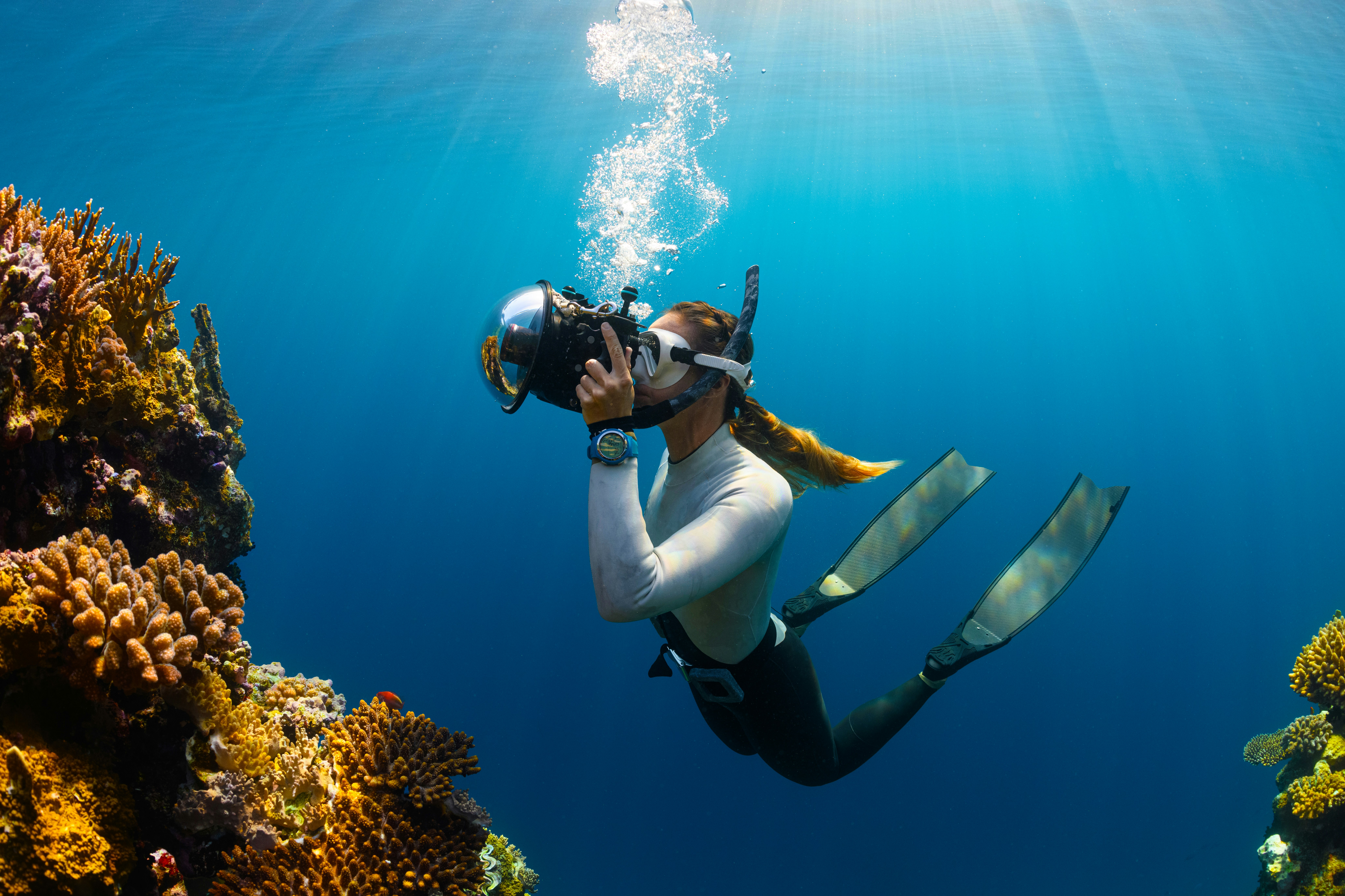
Subfoveal choroid and RPE thickening, which is also observed in pachychoroid pigment epitheliopathy, was identified as a long-term effect of diving in deep divers. Photo: NEOM on Unsplash. |
Diving, an intense activity placing the body under hyperbaric and hyperoxic conditions, has been shown to induce ocular damage. Notably, retinal damage is found to increase with long-term diving. However, very few studies have evaluated the retina in professional divers, and there are no studies comparing the retinas of deep and scuba divers. Researchers in Turkey evaluated the long-term effects of diving on the thickness of retinal layers and retinal anatomy in these groups. Their findings, which were published in Journal of Ophthalmology, revealed that an increased thickening of the subfoveal choroid and retinal pigment epithelium (RPE), resembling pachychoroid pigment epitheliopathy, was detected in deep divers over a long-term duration.
The study included 52 eyes of deep divers who dive to depths of more than 130 feet, 49 eyes of scuba divers who dive up to 130 feet and 66 eyes of the control group, consisting of nondiving but regularly exercising males. Measurements of macular retinal layer thicknesses, peripapillary nerve fiber layer thickness, subfoveal choroidal thickness and peripheral retinal examinations with scleral indentation were performed and statistically compared between the groups.
The mean diving duration was 10.7 years in the deep diver group and 13.9 years in the scuba diver group, with the median diving duration being eight years (two to 30 years) in the deep divers and 12 years (two to 31 years) in the scuba divers. The RPE was statistically significantly thicker in deep divers than in scuba divers and the control group on the 3mm ring of the Early Treatment Diabetic Retinopathy Study grid. Subfoveal choroidal thickness was significantly thicker in deep divers than in scuba divers.
RPE abnormalities showed a significant increase in both the deep and scuba diver groups. Besides the statistically significant increase in RPE layer thickness in deep divers, other RPE abnormalities, such as RPE spikes and pachydrusen, were also significantly more frequent in the deep divers. These findings suggest pachychoroid pigment epitheliopathy.
When discussing the potential mechanisms that lead to these findings, the researchers highlighted that, while scuba divers inhale through a separate device from the mouth, they exhale into a half-face mask covering the periorbital area and nose. During exhalation, they attempt to create a small amount of positive pressure inside the mask. This positive pressure applied to the orbit in scuba divers may cause flattening of the globe and reduction of the choroidal layer due to this morphological change.
“Deep divers, on the other hand, dive with helmets that cover the entire head and face. Therefore, the pressure around the eyes and cranium is equal in deep divers,” the study authors wrote in their paper. “Our hypothesis is that deep divers experience intracranial and periorbital venous congestion originating from the neck, which results in choroidal thickening.”
Because RPE abnormalities were a common finding in both diver groups, this may indicate that, in addition to venous congestion and pressure imbalances, the involvement of endothelial dysfunction and other related pathophysiological mechanisms related to diving are at play.
| Click here for journal source. |
Demir N, Kayhan B, Acar M, et al. Retinal layer and choroidal changes in deep and scuba divers: evidence of pachychoroid spectrum-like findings. J Ophthalmol. November 15, 2024. [Epub ahead of print]. |


