 |
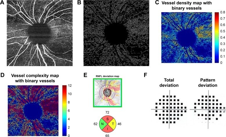
| These OCT-A images from a different study show the process of vascular skeleton density measurement in glaucoma and correlation with visual field results: (A) En face image. (B) Skeletonized vessel image with large vessels removed. (C) Vessel density map with binary vessels showing areas of higher vessel density in warmer colors. (D) Vessel complexity map with binary vessels showing areas of greater vessel branching in warmer colors. (E) OCT RNFL deviation map and RNFL thickness by quadrant. (F) Probability total and pattern deviation maps. Photo: Richter GM, et al. Clin Ophthalmol 8 Nov 2018:2285-2296. Click image to enlarge. |
Research increasingly demonstrates the vascular underpinnings of glaucomatous development. The idea that vasculature is of clinical importance as it relates to glaucoma is gaining favor as a result, given that the condition is linked to factors like blood pressure, intraocular pressure, vascular constriction, cardiovascular diseases and ocular blood circulation.
Due to these growing associations, researchers from China recently sought to investigate the association between retinal microvasculature parameters and glaucoma in a prospective study making use of a large public database. Included were 41,632 participants of the UK Biobank without prior glaucoma and who also had fundus images. To quantify parameters of retinal vascular skeleton density (VSD) and fractal dimension (FD)—two OCT-A metrics mostly used in research settings rather than clinics—the investigators used an AI-based system to segment retinal vascular structures for quantification. Propensity score matching was also employed to pair glaucoma patients with healthy controls. Results were recently published in Ophthalmology Science.
Of the large set of individuals included, 482 glaucoma cases were recorded over a median follow-up of 11 years. After analysis, the researchers found that cases of glaucoma had negative associations with arteriolar VSD (hazard ratio: 0.24), venular VSD (HR: 0.34), arteriolar FD (HR: 0.24) and venular FD (HR: 0.31). Additionally, subgroup analysis with covariates revealed that those aged 60 and older, non-smokers, moderate alcohol consumers and those with hypertension and myopia exhibited p-values consistently below 0.05 both pre- and post-matching.
Overall, the authors indicate in their paper on the work that “retinal microvascular characteristics could play a significant role in the pathogenesis of glaucoma, thereby opening up the prospect of utilizing retinal fundus images to develop a deep-learning predictive model to assess the risk of glaucoma.”
In the paper, the researchers elaborate on the importance of VSD, which is often used to measure capillary density, determined by the proportion of blood vessel length to the overall area in the skeletonized vessel map. As they explain, assessing retinal vasculature in the macula may enhance glaucomatous changes’ detection; this is because of retinal ganglion cells high metabolic demands and reliance on local capillary networks. What’s more, decreased vessel density in eyes with glaucoma may suggest reduced metabolic demand for retinal ganglion cells.
As well as retinal vessel density, FD of retinal vessels was also linked to glaucoma presence. FD clarifies the extent to which a pattern occupies areas in two dimensions, making it a comprehensive metric encapsulating the whole branching structure within the retinal vascular network. The depleted FD of retinal vasculature seen in glaucomatous individuals of this study indicated potential compromise in vascular circulation optimization; this is due to alterations of the geometric pattern being suggestive of deviating from ideal structure and function of microcirculation, thus resulting in less efficiency and impaired circulatory transport.
The authors also explain that static characteristics of retinal microcirculation may be related to vascular events in glaucoma. As such, they believe that “studying early changes in retinal microvascular parameters is clinically valuable for diagnosing glaucoma linked to vascular factors.”
| Click here for journal source. |
Chen Q, Miao S, Jiang Y, et al. Associations of retinal microvascular density and fractal dimension with glaucoma: a prospective study from UK Biobank. Ophthalmol Sci. November 27, 2024. [Epub ahead of print]. |