 |
|
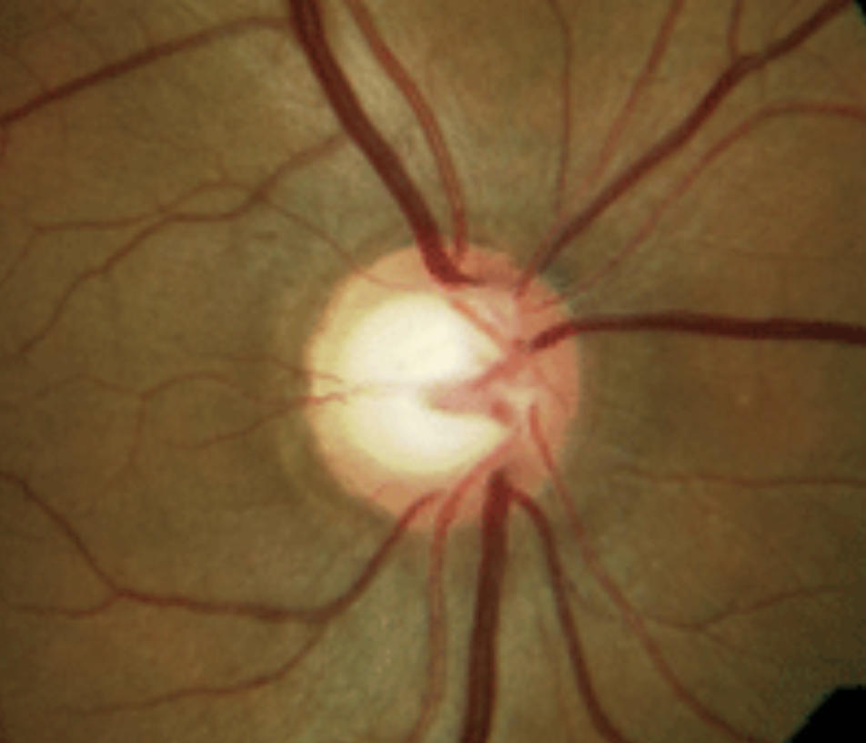
Eyes with physiological cupping only had a weak positive correlation between retinal nerve fiber layer and RPC VD in the superior quadrant, but normal eyes saw moderate positive correlations. Photo: Michael Chaglasian, OD. Click image to enlarge. |
Determining who is glaucoma suspect vs. who actually has the disease can be difficult. This is true for patients with physiological cupping and requires a thorough evaluation to resolve uncertainty. Using OCT-angiography (OCT-A), researchers conducted a recently published study to distinguish between physiological and glaucomatous cupping via radial peripapillary capillaries vessel density (RPC VD).
Included in the investigation were 98 eyes of 98 patients who were then subdivided into three groups: group 1, with 30 eyes, that had primary open angle glaucoma; group 2, with 28 normal eyes but with physiological cupping; and group 3, with 40 age-matched normal eyes.
It was found upon analysis that group 1 had significantly lower RPC VD than the other two groups, and no significant differences were observed between group 2 and 3. In the glaucomatous eyes, moderate negative correlations were observed between both cup-to-disc vertical and horizontal ratios, as well as RPC VD across the entire image and its four individual quadrants.
In light of these findings, the researchers wrote in their paper for Journal of Glaucoma that “our findings indicate that cupping does not influence the decrease in RPC VD, which is primarily linked to glaucomatous damage.”
Further discussion revealed that a difference was seen in ganglion cell complex and retinal nerve fiber layer thickness. Glaucomatous eyes had the thinnest when compared with both other groups.
The investigators also confirmed that those with physiological cupping had no progressive signs of damage, due to lack of consensus on definitive clinical diagnostic or follow-up guidelines for these individuals. This was demonstrated through normal visual field testing and stable, normal within-range measurements for the retinal nerve fiber layer on at least two OCT scans taken six months apart or longer.
Growing evidence supports a hemodynamic effect of glaucomatous optic neuropathy, but with controversy surrounding vascular attenuation and capillary dropout seen in glaucomatous eyes, as to whether they develop subsequent ganglion cell complex loss and less blood supply demand, or if they precede ganglion cell complex loss and instead have a causative role. Since limited data exists currently for RPC VD in eyes with physiological cupping, the authors wanted to see if RPC VD could contribute additional distinguishing factors between physiological cupping vs. glaucoma.
This idea proved to be useful, as based on their results, “clinically, incorporating OCT-A into the diagnostic process can enhance the accuracy of glaucoma diagnosis and reduce the risk of overdiagnosis and overtreatment by clearly differentiating between benign physiological cupping and pathological glaucomatous changes.”
| Click here for journal source. |
Ashour DM, Madkour NS, Ebeid WM, Mahmoud RA. Peripapillary vascular density differentiates glaucomatous cupping from physiological cupping using optical coherence tomography angiography. J Glaucoma. December 16, 2024. [Epub ahead of print]. |
